Exercise 1
Vocabulary ボキャブラリー
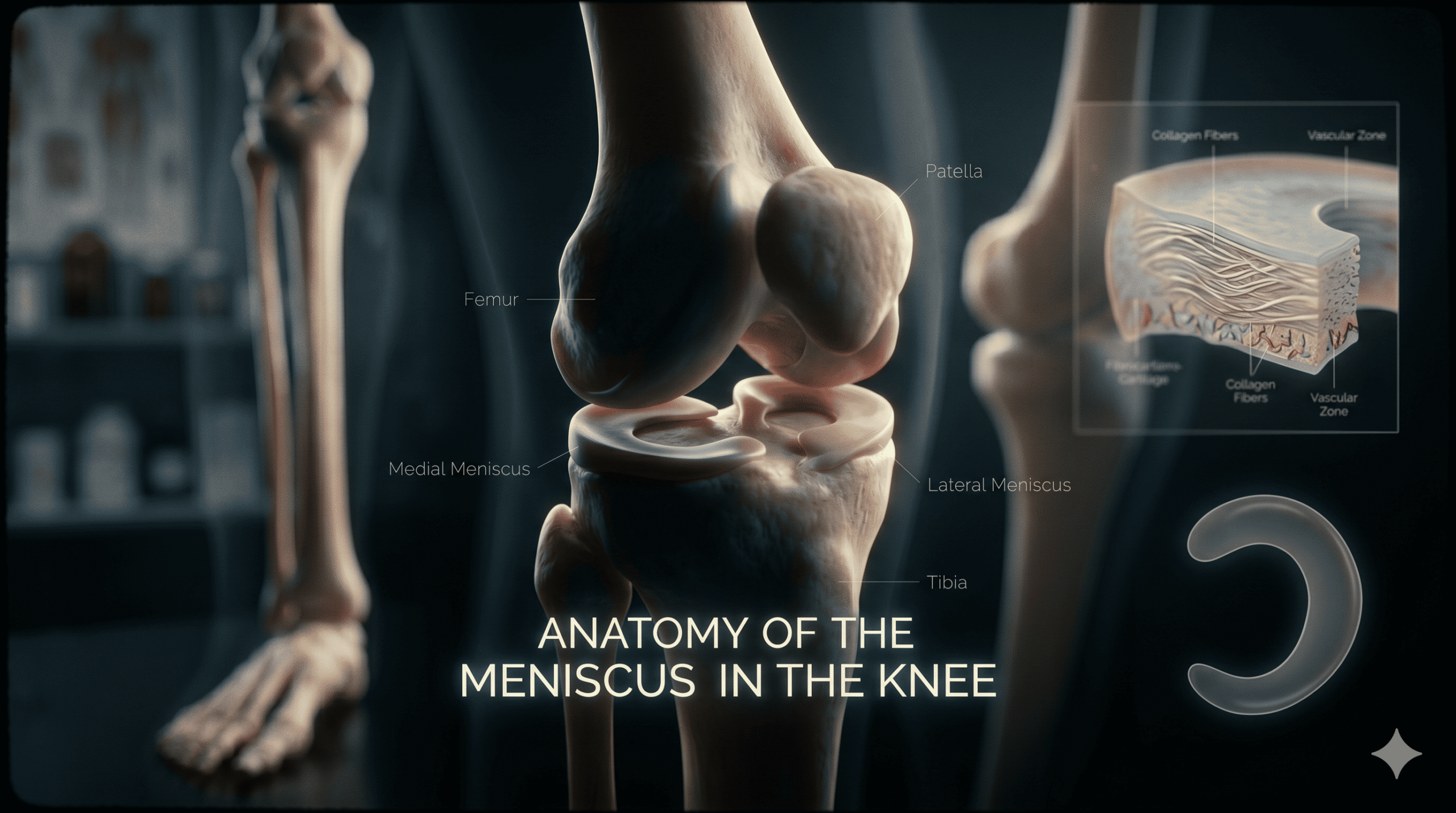
(/məˈnɪskəs/)
名詞
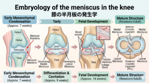
半月板
A C-shaped piece of fibrocartilage in the knee joint that acts as a shock absorber.
Example: The medial meniscus is longer and less mobile than the lateral meniscus.
内側半月板は外側半月板よりも長く、可動性が低い。
(/pɒˈstɪəriər/)
形容詞
後方の
Located at or toward the back of the body or a structure.
Example: Zone 4, located near the posterior horn, is a common site for meniscal tears.
後節の近くに位置するゾーン4は、半月板損傷の一般的な部位である。
(/kəˈlætərəl ˈlɪɡəmənt/)
名詞
側副靭帯
A band of fibrous tissue that connects bones and stabilizes the sides of a joint.
Example: The medial meniscus is stabilized in part by the medial collateral ligament.
内側半月板は、一部は内側側副靭帯によって安定化されている。
(/ˌfaɪbroʊˌkɑːrtɪˈlædʒɪnəs/)
形容詞
線維軟骨性の
Relating to or composed of fibrocartilage, a tough and dense cartilage tissue.
Example: The root attachments of the meniscus feature fibrocartilaginous structures to withstand joint stress.
半月板の付着部根元は、関節のストレスに耐えるために線維軟骨性の構造を特徴としている。
(/ɪkˈstruːʒən/)
名詞
逸脱、押し出し
The state of being pushed or displaced out of its normal anatomical position.
Example: Proper anchoring of the meniscus roots helps prevent meniscal extrusion during joint loading.
半月板根元が適切に固定されることで、関節荷重時の半月板逸脱を防ぐことができる。
Exercise 2
Medical Article
※AI音声
The medial meniscus measures approximately 45.7 mm in length and 27.4 mm in width.
Its width decreases from posterior to anterior and has been reported to range from 12.6 to 17.4 mm in the posterior third, 9.3 to 12.2 mm in the middle third, and 7.6 to 9.0 mm in the anterior third of the medial meniscus.
The thickness of the medial meniscus is relatively consistent from anterior to posterior and ranges from 5.2 to 6.9 mm in thickness over the entire meniscus.
The medial meniscus can also be divided into five anteroposterior zones (Fig.1).
This includes the anterior root attachment (zone 1), the anteromedial zone between the posterior border of the anterior root and the anterior border of the superficial medial collateral ligament (zone 2A and 2B), the portion of the meniscus that is adjacent to the superficial medial collateral ligament (zone 3), the posterior horn (zone 4), and the posterior root (zone 5).
Of clinical note, zone 4 is the most common location for meniscal tears and is the location where meniscal repair is most commonly performed.
Anatomic studies have demonstrated that the medial meniscus covers between 51 and 74% of the medial tibial plateau surface area.
A study by Bloecker et al.
attempted to examine this property using MRI and found that the medial meniscus covered 50% of the medial tibial plateau.
The lateral meniscus measures approximately 35.7 mm in length and 29.3 mm in width.
Unlike the medial meniscus, the width of the lateral meniscus is relatively consistent across the entire structure for a given specimen.
It has been reported to range from 9.8 to 12.0 mm in the posterior third, 10.0 to 12.5 mm in the middle third, and 10.0 to 11.9 mm in the anterior third of the meniscus.
The lateral meniscus is thinner on the anterior third, where it ranges from 3.8 to 4.73 mm in thickness.
The thicker middle third and posterior third range from 5.9 to 6.5 mm and 5.3 to 6.2 mm in thickness, respectively.
The lateral meniscus can be classified into six zones based on anteroposterior location (Fig. 1).
These include the anterior root (zone 1), the anterolateral zone between the anterior root and the anterior border of the popliteal hiatus (zones 2A and 2B), the popliteal hiatus (zone 3), the posteroinferior popliteomeniscal fascicle (zone 4), the ligamentous zone (zone 5), and the posterior root (zone 6).
Anatomic studies have demonstrated that the lateral meniscus covers between 75 and 93% of the lateral tibial plateau surface area.
The MRI study by Bloecker et al.
found that the lateral meniscus covered 59% of the lateral tibial plateau. The menisci are attached to the tibial plateau via their anterior and posterior roots and are stabilized by the medial collateral ligament, the transverse ligament, the meniscotibial ligaments, and the meniscofemoral ligaments.
The roots of the meniscus are ligamentous-like structures with fibrocartilaginous entheses.
They are essential to function as they anchor the meniscus to convert axial loads into hoop stresses and prevent extrusion during joint loading.
The meniscotibial ligament anchors the entire outer margin of the medial meniscus to the medial tibial plateau.
The medial meniscus also has attachments to the posterior oblique ligament and the posteromedial capsule.
The majority of the outer border of the lateral meniscus is attached to the lateral tibial plateau by the meniscotibial ligament.
The lateral meniscotibial ligament is thinner and more elastic than the medial meniscotibial ligament.
Additionally, the crossing of the popliteus tendon partially disrupts this circumferential attachment.
Together, these properties allow the lateral meniscus to have increased translation and mobility relative to the more static medial meniscus.
内側半月板は、長さ約45.7 mm、幅約27.4 mmである。
その幅は後方から前方にかけて減少し、内側半月板の後1/3(後節)で12.6〜17.4 mm、中1/3(中節)で9.3〜12.2 mm、前1/3(前節)で7.6〜9.0 mmの範囲であると報告されている。
内側半月板の厚さは前方から後方にかけて比較的均一であり、半月板全体で5.2〜6.9 mmの範囲である。
内側半月板は、前後の位置に基づいて5つのゾーンに分けることもできる(図1)。
これには、前節付着部(ゾーン1)、前節付着部の後縁から浅内側側副靭帯の前縁までの間の前内側ゾーン(ゾーン2Aおよび2B)、浅内側側副靭帯に隣接する半月板の部分(ゾーン3)、後節(ゾーン4)、および後節付着部(ゾーン5)が含まれる。
臨床的に注目すべき点として、ゾーン4は半月板損傷の最も一般的な部位であり、半月板修復術が最も頻繁に行われる場所でもある。
解剖学的研究により、内側半月板は内側脛骨高原(脛骨顆間区)の表面積の51〜74%を覆っていることが示されている。
Bloeckerらによる研究では、MRIを用いてこの特性を調査しようと試み、内側半月板が内側脛骨高原の50%を覆っていることを突き止めた。
外側半月板は、長さ約35.7 mm、幅約29.3 mmである。
内側半月板とは異なり、外側半月板の幅は、特定の標本において構造全体を通じて比較的均一である。
外側半月板の後1/3で9.8〜12.0 mm、中1/3で10.0〜12.5 mm、前1/3で10.0〜11.9 mmの範囲であると報告されている。
外側半月板は前1/3でより薄く、厚さは3.8〜4.73 mmの範囲である。
より厚い中1/3および後1/3の厚さは、それぞれ5.9〜6.5 mmおよび5.3〜6.2 mmの範囲である。
外側半月板は、前後の位置に基づいて6つのゾーンに分類できる(図1)。
これらには、前節付着部(ゾーン1)、前節付着部から膕筋裂孔の前縁までの間の前外側ゾーン(ゾーン2Aおよび2B)、膕筋裂孔(ゾーン3)、後下膕半月束(ゾーン4)、靭帯ゾーン(ゾーン5)、および後節付着部(ゾーン6)が含まれる。
解剖学的研究により、外側半月板は外側脛骨高原表面積の75〜93%を覆っていることが示されている。
BloeckerらによるMRI研究では、外側半月板が外側脛骨高原の59%を覆っていることがわかった。
半月板は、前節および後節の付着部を介して脛骨高原に付着しており、内側側副靭帯、横靭帯、半月脛骨靭帯、および半月大腿靭帯によって安定化されている。
半月板の付着部は、線維軟骨性のエンテシス(靭帯付着部)を持つ靭帯様の構造である。
これらは、半月板を固定して軸方向の荷重をフープストレス(周方向応力)に変換し、関節への荷重負荷時に逸脱(押し出し)を防ぐために不可欠な機能を持っている。
半月脛骨靭帯は、内側半月板の外縁全体を内側脛骨高原に固定している。
内側半月板は、後斜靭帯および後内側関節包にも付着部を持っている。
外側半月板の外縁の大部分は、半月脛骨靭帯によって外側脛骨高原に付着している。
外側半月脛骨靭帯は、内側半月脛骨靭帯よりも薄く、伸縮性に富んでいる。
さらに、膝窩筋腱の交差がこの周方向の付着を部分的に途切れさせている。
これらの性質が組み合わさることで、外側半月板は、より静的な内側半月板と比較して、可動性と移動(トランスレーション)が大きくなっている。
Fig. 1
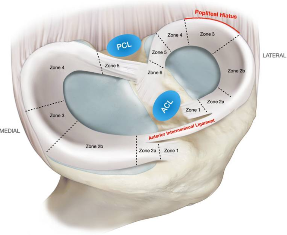
Reference: Enzo S Mameri, Suhas P Dasari, Luc M Fortier, Fernando Gómez Verdejo, Safa Gursoy, Adam B Yanke, Jorge Chahla
Review of Meniscus Anatomy and Biomechanics
Published: 10 August 2022 Volume 15, pages 323–335 (2022) Current Reviews in Musculoskeletal Medicine PMCID: PMC9463428 PMID: 35947336
Quoted from Figure 2
Exercise 3
Discussion ディスカッション
Q: Which zone of the medial meniscus is the most common location for tears?
内側半月板のどのゾーンが、最も一般的に損傷が起こる部位ですか?
Q: What percentage of the medial tibial plateau surface area does the medial meniscus cover according to anatomic studies?
解剖学的研究によると、内側半月板は内側脛骨プラトーの表面積の何パーセントを覆っていますか?
Q: How does the width of the lateral meniscus differ from that of the medial meniscus?
外側半月板の幅は、内側半月板の幅とどのように異なりますか?
Q: What is the primary function of the meniscus roots during joint loading?
関節への荷重負荷時における半月板根元(付着部)の主な機能は何ですか?
Q: Why does the lateral meniscus have increased mobility compared to the medial meniscus?
なぜ外側半月板は内側半月板に比べて可動性が高いのですか?
Exercise 4
Further Discussion 一歩踏み込んだディスカッション
Q: In your clinical experience, what are the most common challenges patients face when recovering from a zone 4 meniscal tear?
あなたの臨床経験において、ゾーン4の半月板損傷から回復する際に患者が直面する最も一般的な課題は何ですか?
Q: How do you decide between recommending conservative physical therapy versus referring a patient for meniscal repair surgery?
保存的な理学療法を勧めるか、それとも半月板修復術のために手術を勧めるかは、どのように判断しますか?
Q: What specific exercise progressions do you find most effective for stabilizing the knee joint after a meniscal injury?
半月板損傷後の膝関節を安定させるために、どのような具体的な段階的エクササイズが最も効果的だと考えていますか?
Q: Given the higher mobility of the lateral meniscus, how does your rehabilitation approach differ for lateral versus medial meniscal tears?
外側半月板の可動性が高いことを考慮すると、外側半月板損傷と内側半月板損傷のリハビリテーションアプローチはどのように異なりますか?
Q: How do you educate patients about the risk of long-term joint degeneration (such as osteoarthritis) following a meniscal extrusion?
半月板逸脱の後に起こる長期的な関節変形(変形性膝関節症など)のリスクについて、患者にどのように説明しますか?
Reference
Review of Meniscus Anatomy and Biomechanics
Enzo S Mameri, Suhas P Dasari, Luc M Fortier, Fernando Gómez Verdejo, Safa Gursoy, Adam B Yanke, Jorge Chahla
URL:https://pmc.ncbi.nlm.nih.gov/articles/PMC9463428/