Exercise 1
Vocabulary ボキャブラリー
名詞
扁桃体
a roughly almond-shaped mass of gray matter inside each cerebral hemisphere, involved with the experiencing of emotions
Example: The amygdala plays a central role in processing fear and anxiety.
扁桃体は、恐怖と不安の処理において中心的な役割を果たします。
名詞
海馬
the elongated ridges on the floor of each lateral ventricle of the brain, thought to be the center of emotion, memory, and the autonomic nervous system
Example: Reduced activity in the hippocampus was negatively correlated with illness duration.
海馬における活動の低下は、罹病期間と負の相関を示しました。
名詞
楔前部
a part of the superior parietal lobule hidden in the medial longitudinal fissure between the two cerebral hemispheres
Example: Acupuncture reduced synchronization values in several brain regions, including the precuneus.
鍼治療は、楔前部を含む複数の脳領域において同期値を低下させました。
名詞
グルタミン酸
a salt or ester of glutamic acid, functioning as a primary excitatory neurotransmitter in the central nervous system
Example: Excessive excitatory signaling in the brain involving glutamate can lead to neuronal hyperexcitability.
脳内のグルタミン酸が関与する過剰な興奮性シグナル伝達は、神経の過剰興奮を引き起こす可能性があります。
名詞
神経伝達物質
a chemical substance that is released at the end of a nerve fiber by the arrival of a nerve impulse and, by diffusing across the synapse or junction, causes the transfer of the impulse to another nerve fiber, a muscle fiber, or some other structure
Example: Acupuncture modulates neurotransmitter-related signaling within brain regions.
鍼治療は、脳領域内の神経伝達物質に関連するシグナル伝達を調節します。
Exercise 2
Medical Article
※AI音声
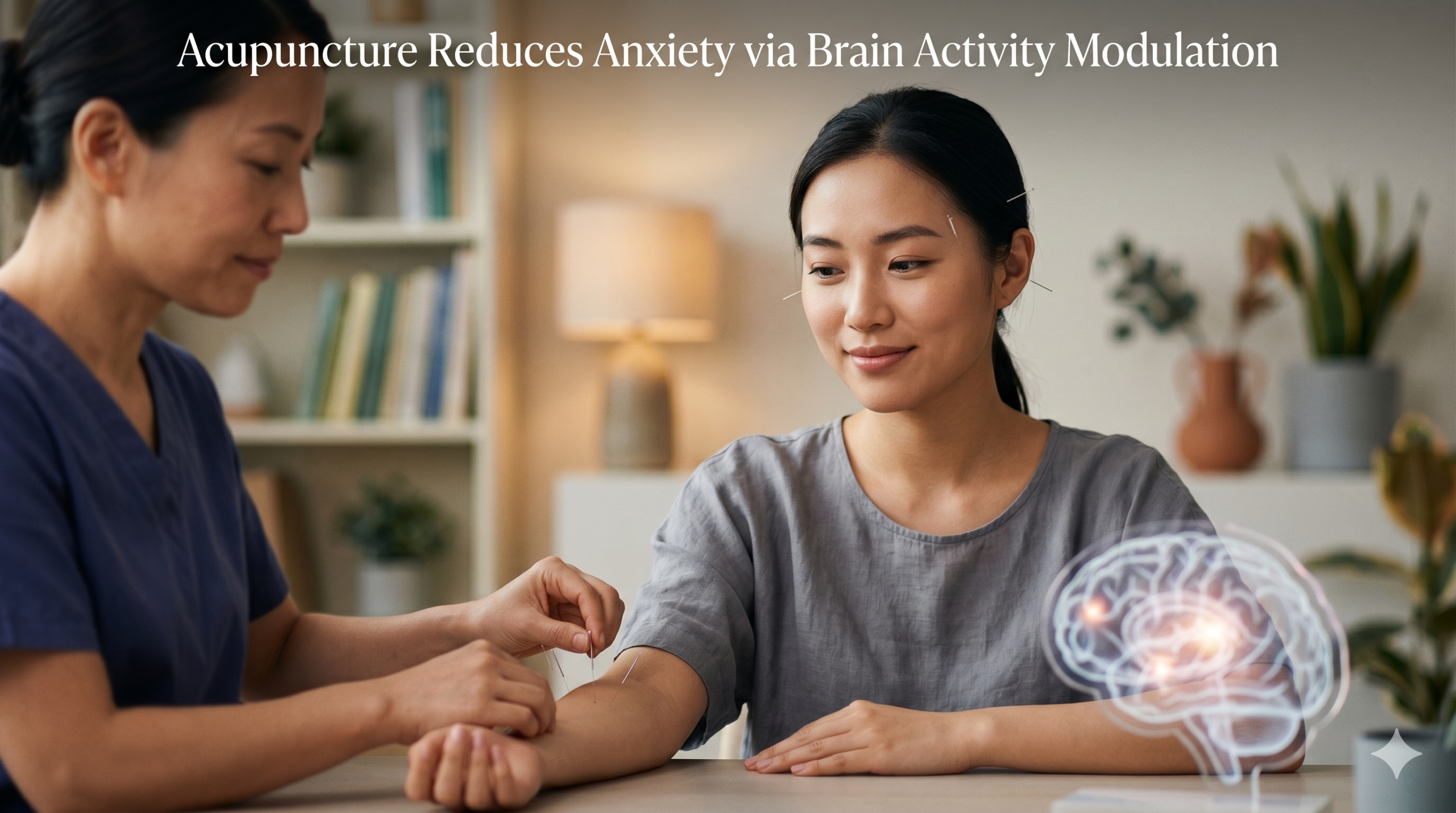
Acupuncture reduces anxiety symptoms in patients with generalized anxiety disorder (GAD), with measurable improvements on standardized clinical assessments and objective changes in brain activity.
Researchers report that patients receiving acupuncture experienced significant reductions in anxiety severity compared with a control group, alongside measurable changes in how the brain regulates activity at rest.
In a controlled clinical study conducted at the First Affiliated Hospital of Hainan Medical University, 70 patients meeting DSM-5 criteria for GAD were randomly assigned to an acupuncture group or a wait-list control group.
After a 4-week treatment period, the acupuncture group demonstrated a significant reduction in anxiety severity as measured by the Hamilton Anxiety Rating Scale (HAMA), while the wait-list group showed no statistically significant change.
The HAMA is a clinician-administered scale that evaluates both psychological symptoms, such as excessive worry and tension, and physical symptoms, including restlessness and somatic discomfort.
To examine how acupuncture affects the brain, researchers used resting-state functional magnetic resonance imaging (fMRI). [Image of fMRI scan of the brain]
This imaging method measures brain activity indirectly by detecting changes in blood oxygen levels, which reflect underlying neural activity.
Because the scan is performed while the patient is not engaged in any specific task, it captures baseline patterns of brain function and how different regions maintain activity and coordination under resting conditions.
The analysis focused on regional homogeneity (ReHo), a measure of how synchronized neighboring brain cells are within a small, localized area.ReHo measures how similar the activity is among neighboring voxels — the smallest measurable units in fMRI data — each of which captures the collective activity of a large population of neurons.
When neurons in a region are firing in a highly coordinated way, ReHo values are higher, indicating tightly coupled activity.
When this synchronization decreases, ReHo values decline, reflecting less coordinated local activity.
In the context of anxiety disorders, increased synchronization in certain brain regions can reflect heightened or persistent activation of circuits involved in emotional processing.
The study found that acupuncture reduced ReHo values in several brain regions associated with emotional regulation and stress processing.
These included the amygdala, hippocampus, anterior and posterior cingulate cortices, putamen, and precuneus.
The amygdala, which plays a central role in processing fear and anxiety, showed changes that were directly associated with clinical outcomes.
Specifically, reductions in ReHo within the amygdala were positively correlated with decreases in HAMA scores, indicating that patients with greater reductions in local neural synchronization in this region experienced greater improvement in anxiety symptoms.
In addition, reduced ReHo in the hippocampus was negatively correlated with illness duration, suggesting a relationship between hippocampal activity and the chronicity of anxiety.
To clarify the biological meaning of these findings, the authors examined neurotransmitter-related activity in parallel animal experiments.
They note that excessive excitatory signaling in the brain—particularly involving glutamate, a primary excitatory neurotransmitter—can lead to neuronal hyperexcitability.
This means that neurons fire more easily and more frequently than normal.
Such heightened excitability in regions like the amygdala is associated with increased synchronization of neural activity, which is reflected as elevated ReHo on fMRI.
The reduction in ReHo observed after acupuncture is therefore consistent with a decrease in this heightened neural activity, indicating a shift toward less synchronized firing in these regions.
The acupuncture protocol used in the clinical portion of the study was standardized.
Acupoints included Baihui (GV20), Neiguan (PC6), Shenmen (HT7), and Taichong (LV3). Disposable sterile needles (0.25 × 25 mm and 0.25 × 40 mm) were inserted bilaterally.
GV20 was inserted transversely, while PC6, HT7, and LV3 were inserted perpendicularly. Needle manipulation consisted of lifting, thrusting, and twisting techniques.
Each treatment session lasted 30 minutes and was administered 4–5 times per week over 4 weeks, for a total of 16–20 treatments.
[1] A parallel lab experiment was used to confirm the human clinical findings.
To further investigate mechanisms, the study incorporated a chronic unpredictable stress (CUS) rat model, which induces anxiety-like behaviors through repeated exposure to varying stressors.
Electroacupuncture (EA) was applied at the same acupoints using anatomically scaled parameters.
Needles (0.25 mm diameter) were inserted to depths of approximately 5 mm at GV20 and LV3 and 2–3 mm at PC6 and HT7.
Electrical stimulation was delivered using a Han’s Acupoint Nerve Stimulator at alternating frequencies of 2/15 Hz for 25 minutes once daily over 21 consecutive days, with intensity adjusted to produce mild limb tremors.
Behavioral testing demonstrated that EA improved anxiety-like responses.
In the elevated plus maze, which assesses avoidance of open spaces, EA increased both open arm entry percentage and open arm time percentage compared with the stress model group.
In the open field test, which evaluates movement and exploratory behavior, EA reduced total travel distance and average speed and increased time spent in the central area.
These findings indicate reduced anxiety-like behavior following electroacupuncture.
No comparable improvements were observed in the sham acupuncture group.
Molecular analyses examined neurotransmitter-related signaling within the medial prefrontal cortex (mPFC) and amygdala.
In the stress model, expression of insulin-like growth factor-1 (IGF-1) in the mPFC was reduced, while expression of NR2B and GluR2 in the amygdala was increased.
NR2B is a subunit of the NMDA receptor, and GluR2 is part of the AMPA receptor complex; both are involved in excitatory neurotransmission.
Following electroacupuncture, IGF-1 expression increased in both the mPFC and amygdala, while NR2B, GluR1, and GluR2 expression in the amygdala were reduced.
These changes were confirmed at both gene and protein levels using quantitative PCR and Western blot analysis.
Immunofluorescence staining further demonstrated reduced NR2B expression in the amygdala.
Additional findings showed that electroacupuncture improved physiological measures under stress conditions.
Body weight, which decreased during chronic stress exposure, increased following electroacupuncture treatment, while no significant changes were observed in the sham group.
Taken together, the clinical, imaging, and molecular findings indicate that acupuncture reduces anxiety symptoms, alters baseline brain activity, and modulates neurotransmitter-related signaling within brain regions involved in emotional regulation.
The results identify changes in the medial prefrontal cortex–amygdala pathway as a central feature associated with these effects.
鍼治療は、全般性不安障害(GAD)患者の不安症状を軽減し、標準化された臨床評価における測定可能な改善と、脳活動の客観的な変化をもたらします。
研究者らは、鍼治療を受けた患者は対照群と比較して不安の重症度が有意に低下し、それとともに安静時の脳の活動調節方法に測定可能な変化が生じたと報告しています。
海南医科大学第一附属病院で実施された対照臨床試験では、GADのDSM-5基準を満たす70名の患者が、鍼治療群または待機的対照群のいずれかに無作為に割り当てられました。
4週間の治療期間後、鍼治療群はハミルトン不安評価尺度(HAMA)で測定された不安の重症度において有意な低下を示しましたが、待機的対照群では統計的に有意な変化は見られませんでした。
HAMAは臨床医が評価する尺度であり、過度な心配や緊張などの心理的症状と、落ち着きのなさや身体的不快感などの身体的症状の両方を評価するものです。
鍼治療が脳に与える影響を調べるため、研究者らは安静時機能的磁気共鳴画像法(fMRI)を使用しました。[脳のfMRIスキャン画像]
この画像化手法は、基礎となる神経活動を反映する血中酸素濃度の変化を検出することで、脳活動を間接的に測定します。
このスキャンは患者が特定の課題(タスク)を行っていない状態で行われるため、脳機能のベースラインのパターンや、安静時において異なる脳領域がどのように活動と協調性を維持しているかを捉えることができます。
解析では、狭い局所的な領域内で隣接する脳細胞がどの程度同期しているかを示す指標である局所的一貫性(ReHo:Regional Homogeneity)に焦点を当てました。
ReHoは、隣接するボクセル(fMRIデータにおいて測定可能な最小単位で、それぞれが多数の神経細胞集団の集合的な活動を捉える)間で活動がどれだけ類似しているかを測定します。
ある領域内の神経細胞が高度に協調して発火している場合、ReHo値は高くなり、活動が密接に結びついていることを示します。
この同期性が低下するとReHo値も低下し、局所的な活動の協調性が低くなっていることを反映します。
不安障害の文脈においては、特定の脳領域における同期性の増加は、感情処理に関与する神経回路の過剰または持続的な活性化を反映している可能性があります。
本研究では、鍼治療が感情調節とストレス処理に関連するいくつかの脳領域でReHo値を低下させることがわかりました。
これらには、扁桃体、海馬、前部および後部帯状皮質、被殻、楔前部(けつぜんぶ)が含まれます。恐怖や不安の処理において中心的な役割を果たす扁桃体は、臨床的転帰と直接的に関連する変化を示しました。
具体的には、扁桃体内のReHoの低下はHAMAスコアの低下と正の相関を示しました。
これは、この領域における局所的な神経同期の低下が大きい患者ほど、不安症状の改善が大きいことを示しています。
さらに、海馬におけるReHoの低下は罹病期間と負の相関を示しており、海馬の活動と不安の慢性化との間に関連性があることが示唆されました。
これらの知見の生物学的な意味を明らかにするため、著者らは並行して行われた動物実験で神経伝達物質に関連する活動を調べました。
彼らは、脳内の過剰な興奮性シグナル伝達(特に主要な興奮性神経伝達物質であるグルタミン酸が関与するもの)が神経細胞の過活動(過興奮性)につながる可能性があると指摘しています。
これは、神経細胞が通常よりも容易かつ頻繁に発火することを意味します。
扁桃体のような領域におけるこうした興奮性の高まりは、神経活動の同期性の増加と関連しており、これはfMRI上でのReHoの上昇として反映されます。
したがって、鍼治療後に観察されたReHoの低下は、この高まった神経活動の減少と一致しており、これらの領域において同期性の低い発火へとシフトしたことを示しています。
本研究の臨床部分で使用された鍼治療のプロトコルは標準化されていました。
使用された経穴(ツボ)には、百会(GV20)、内関(PC6)、神門(HT7)、および太衝(LV3)が含まれました。使い捨ての滅菌鍼(0.25 × 25 mm および 0.25 × 40 mm)が両側に刺入されました。
百会(GV20)は横刺(水平方向)で、内関(PC6)、神門(HT7)、太衝(LV3)は直刺(垂直方向)で刺入されました。運鍼操作には、提挿(上下に動かす)および捻転(回転させる)の技術が含まれました。
各治療セッションは30分間で、4週間にわたり週4〜5回実施され、合計16〜20回の治療が行われました。[1] ヒトでの臨床所見を確認するために、並行して研究室での実験(動物実験)が行われました。
メカニズムをさらに調査するために、この研究では、さまざまなストレッサーへの反復的な曝露によって不安様行動を誘発する慢性予測不能ストレス(CUS)ラットモデルを組み込みました。
同じ経穴に対し、解剖学的にスケールを合わせたパラメータを用いて電気鍼治療(EA)が施されました。鍼(直径0.25 mm)は、GV20とLV3には約5 mmの深さに、PC6とHT7には2〜3 mmの深さに刺入されました。
Han’s Acupoint Nerve Stimulator(経穴神経刺激装置)を使用し、2/15 Hzの交互周波数で25分間、21日間連続して1日1回電気刺激を行い、強度は手足に軽度の震えが生じる程度に調整されました。
行動試験により、電気鍼治療が不安様反応を改善することが実証されました。開けた空間の回避を評価する高架式十字迷路において、電気鍼治療はストレスモデル群と比較して、オープンアーム(壁のない通路)への進入率および滞在時間率の両方を増加させました。
運動や探索行動を評価するオープンフィールド試験において、電気鍼治療は総移動距離と平均速度を減少させ、中央エリアでの滞在時間を増加させました。
これらの結果は、電気鍼治療後に不安様行動が減少したことを示しています。
偽鍼治療(シャム鍼)群では、同等の改善は観察されませんでした。
分子解析では、内側前頭前野(mPFC)および扁桃体内の神経伝達物質に関連するシグナル伝達を調べました。
ストレスモデルでは、mPFCにおけるインスリン様成長因子-1(IGF-1)の発現が減少する一方で、扁桃体におけるNR2BおよびGluR2の発現が増加していました。
NR2BはNMDA受容体のサブユニットであり、GluR2はAMPA受容体複合体の一部です。
どちらも興奮性神経伝達に関与しています。電気鍼治療後、mPFCと扁桃体の両方でIGF-1の発現が増加し、扁桃体におけるNR2B、GluR1、GluR2の発現は減少しました。
これらの変化は、定量的PCRおよびウェスタンブロット解析を用いて、遺伝子およびタンパク質レベルの両方で確認されました。
免疫蛍光染色ではさらに、扁桃体におけるNR2Bの発現低下が実証されました。
追加の知見として、電気鍼治療がストレス条件下での生理学的指標を改善したことが示されました。
慢性的なストレス曝露中に減少した体重は電気鍼治療後に増加しましたが、偽鍼治療群では有意な変化は観察されませんでした。
これらを総合すると、臨床的、画像的、および分子的知見は、鍼治療が不安症状を軽減し、安静時の脳活動を変化させ、感情調節に関与する脳領域内の神経伝達物質関連シグナル伝達を調整することを示しています。
この結果は、内側前頭前野-扁桃体経路における変化が、これらの効果に関連する中心的な特徴であることを特定しています。
Exercise 3
Discussion ディスカッション
Q: What is the primary function of the Hamilton Anxiety Rating Scale (HAMA) as mentioned in the study?
研究で言及されているハミルトン不安評価尺度(HAMA)の主な役割は何ですか?
Q: How does resting-state fMRI indirectly measure brain activity?
安静時fMRIはどのようにして脳活動を間接的に測定しますか?
Q: What does a higher regional homogeneity (ReHo) value indicate about neurons in a specific brain area?
局所領域均一性(ReHo)の高い値は、特定の脳領域内のニューロンについて何を示していますか?
Q: Which specific acupoints were utilized during the human clinical portion of this research?
この研究の人間を対象とした臨床部分では、具体的にどの経穴(ツボ)が使用されましたか?
Q: According to the animal experiments, what effect did electroacupuncture have on NR2B and GluR2 expression in the amygdala?
動物実験によると、電気鍼は扁桃体におけるNR2BとGluR2の発現にどのような影響を与えましたか?
Exercise 4
Further Discussion 一歩踏み込んだディスカッション
Q: How could the objective evidence provided by fMRI change the way traditional therapies like acupuncture are perceived within modern, evidence-based medicine?
fMRIが提供する客観的証拠は、現代の根拠に基づく医療(EBM)の中で、鍼治療のような伝統療法の認識をどのように変える可能性があると思いますか?
Q: In your own clinical experience, how do you manage and treat patients presenting with physical symptoms rooted in psychological stress or anxiety?
あなた自身の臨床経験において、心理的ストレスや不安を根源とする身体症状を訴える患者をどのように管理・治療していますか?
Q: This study used a standardized set of acupoints. How does this standardized protocol compare with the individualized treatments you typically perform in practice?
この研究では標準化された経穴のセットを使用しました。この標準化されたプロトコルは、あなたが通常実践で行っている個別化された治療とどのように異なりますか?
Q: Do you believe the neurobiological mechanisms observed in rat stress models (like CUS) can be reliably extrapolated to understand human anxiety disorders?
ラットのストレスモデル(CUSなど)で観察された神経生物学的メカニズムから、人間の不安障害を理解するために確実に推論できると思いますか?
Q: How might explaining the scientific and neurobiological mechanisms of a treatment to a patient affect their placebo response or overall adherence to the therapy?
治療の科学的および神経生物学的なメカニズムを患者に説明することは、プラセボ反応や治療の全体的な遵守にどのような影響を与える可能性がありますか?
Sourse
Zhao Sun et al., “Acupuncture Modulates Neurotransmitter-Related Molecules in the Amygdala to Ameliorate Generalized Anxiety Disorder,” CNS Neuroscience & Therapeutics, 2026.